
Page 32 - NEWSLETTER_27
P. 32
E.O.E Newsletter | www.huanet.gr
dissections impossible. What was the solution? Using the operation room as an anatomy laboratory and performing dissections in stillborn infants.
The first problem I tackled was bleeding. (1) At that time, Urologists did not want to perform radical prostatectomies because of severe bleeding and I thought that if we were ever going to make these operations successful, we had to eliminate it. Using the operating room, I discovered a common trunk over the urethra and I developed the surgical technique for ligating it. In doing so, we reduced bleeding and we made it possible to perform a better cancer operation in a bloodless field. Figure 1.
All of a sudden, a patient came to see me shortly thereafter, and he told me that he was fully potent. How can that be if the nerves run through the prostate? Where were the nerves? They must be outside the prostate but the answer was not in any anatomy book. (2)
Around that time, I was elected to the American Association of Genitourinary Surgeons, which is a distinguished Urological Association. On the night before the meeting, there was no social event. So, my wife and I left the resort and went downtown to a restaurant. And as we were walking to our table, I saw an old man who looked lonely. For the first and only time in my life, I went up to a total stranger and asked him: “Are you alone? Would you like to join us for dinner?”. He said yes. He was at the same meeting and the concierge sent him to the same restaurant. His name was Pieter Donker. He was the Professor and Chairman at the University of Leiden in the Netherlands and his specialty was neuro-urology. We had a wonderful dinner and I thought that this was the end of it, but four years later, 4,000 miles away, we met again.
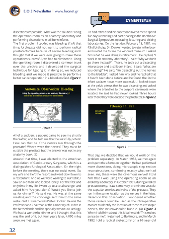
He had retired and his successor invited me to spend five days attending and participating in the Boerhaave Surgical Symposium, operating, lecturing and visiting laboratories. On the last day, February 13, 1981, my 43rd birthday, Dr. Donker wanted to return the favor and invited me to see the windmill museum. I asked him what he was doing in retirement. He replied, “I work in an anatomy laboratory”. I said: “Why we don’t go there instead?”. There, he took out a dissecting microscope and a stillborn infant. I said: “What are you doing?” He said, “I’m dissecting out the nerves to the bladder”. I asked him why and he replied that it hasn’t been done before and he found that in the infant cadaver it was more successful. I looked down at the pelvic plexus that he was dissecting and asked where the branches to the corpora cavernosa were located. He said he had never looked. Three hours later there they were outside the prostate! (3) Figure 2
That day, we decided that we would work on the problem separately. In March 1982, we met again and spent the afternoon together. He had performed more dissections, doing microscopic step section reconstructions, confirming exactly what we had seen. Yes, these were the cavernous nerves! I told him that I was using the operating room as an anatomy laboratory. In October 1981, during a radical prostatectomy, I saw some very prominent vessels: the capsular arteries and veins of the prostate. They ran in the same location as the nerves in the fetus. Based on this observation I wondered whether those vessels could be used as the intraoperative marker to identify the location of these microscopic nerves, the neurovascular bundle. (4) Figure 3 When I told him about this idea he said: “This makes sense to me!”. I returned to Baltimore, and in March 1982 I did a radical cystectomy on a 67-year-old
Anatomical Observations: Bleeding
Using the operating room as an anatomy laboratory, I identified a common trunk over the urethra.
Figure 1
Cavernous nerves
Pelvic Plexus
February 13 1981
Prostate
Figure 2
32
ΣΥΝΈΝΤΈΥΞΗ
